


Retinitis Pigmentosa 79
Patient Information
Characteristics
Background and History
The retina of the eye has many cell types, among them the rods and cones that respond to light and transmit them to the brain. The many biochemical reactions in the rods and cones are under gene control and mutations in these are responsible for a large number of hereditary eye disorders. Among these are a group of conditions called retinitis pigmentosa which resemble each other clinically so closely that gene studies are necessary to distinguish them.
Clinical Correlations
This condition is highly variable with age of onset from childhood through the 6th or 7th decade. Usually the first complaints consist of difficulty seeing at night or in dim light. Vision can remain good until adulthood but most individuals note a slow loss of vision as they get older. With progression, there is often a narrowing of side vision known as peripheral vision constriction. Color vision perception may also deteriorate. A few patients report abnormal sensitivity to light.
The changes in the retina are typical for retinitis pigmentosa with characteristic pigment clumping, narrowing of retinal blood vessels, paleness of the optic nerve, and general patchy wasting of the retina.
Genetics
Inheritance
This is an autosomal dominant disorder that has a vertical transmission pattern. That is, the condition is transmitted directly from an affected parent to each child with a 50% probability.
Pedigree
Autosomal dominant
Autosomal dominant disorders require only one mutation for the disease to be expressed. Since an affected parent has two chromosomes, only one of which has the mutant gene, parents can expect that half (50%) of their children will receive that one and inherit the disease. It is common for individuals that inherit the mutation, however, to not have evidence of the disease (nonpenetrance).
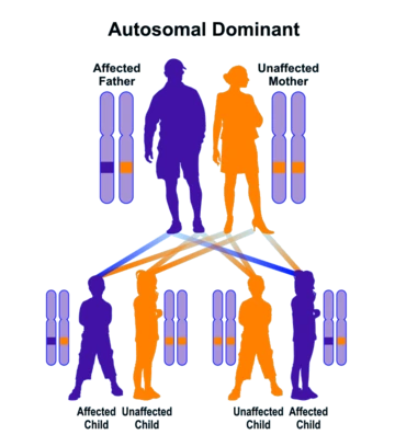
Autosomal dominant inheritance leads to a vertical pattern of transmission
Diagnosis and Prognosis
The diagnosis is made by direct examination of the retina and requires a medical eye doctor: an ophthalmologist. Often special testing such as visual fields and an ERG (electroretinogram) is done. This, however, can only detect the category of disease and gene testing is necessary to determine which condition is actually present.
This condition has no impact on health outside of the eye and lifespan should be normal. The actual visual acuity is highly variable and unpredictable. No treatment has been reported but low vision aids could be helpful especially for near or reading vision.
Printer Friendly Version: Ctrl/Cmd+P